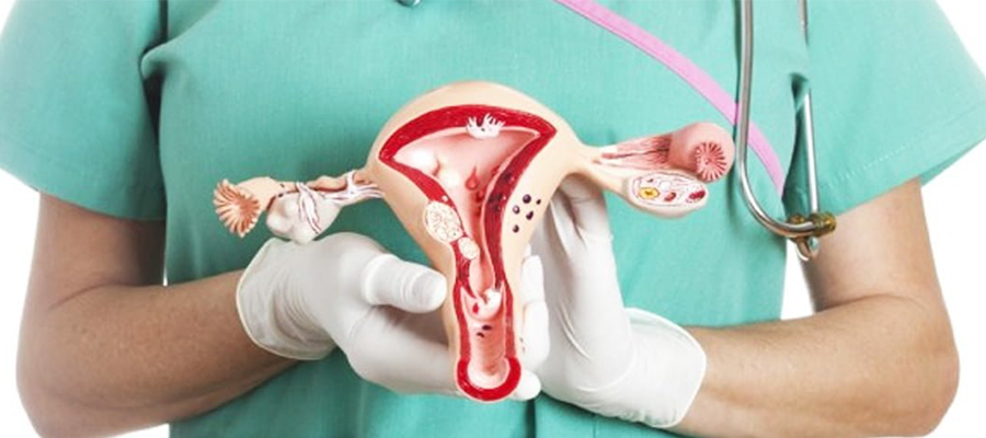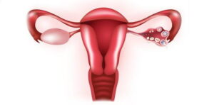
Benign tumors originating from the smooth muscle cells in the myometrium, which we call the uterine wall. myoma uteri It is called. These are mostly seen in women of reproductive age. Rarely, these tumors are found in young girls. Myomas are classified according to their location. Myomas that settle towards the endometrium layer, which causes bleeding in the uterus, are called submucous myomas. Myomas that are in the wall layer are called intramural myomas, and myomas that grow outside the uterus are called subserous myomas.
The reason for this classification is that myomas cause symptoms according to their location. Submucous myomas are more common with prolonged and increased bleeding. Likewise, there is increased bleeding in intramural myomas. Subserous myomas generally do not cause many complaints. Myomas can range from a few millimeters to tens of centimeters. When myomas grow excessively, they can cause many problems.
myoma uteriIt tends to be more common in women who start menstruating early, have never had children, are obese, have a family history of fibroids, and eat a lot of red meat. It is less common in women who have given birth and who exercise regularly.
What Causes Myomas?
First Menstrual Period
In women who have their first menstrual period earlier than normal, the uterus myoma uteri is more likely to happen. This is because the woman is exposed to estrogen for a longer period of time.
Familial Predisposition
Miyomların ortalama %50’sinin ailesel yatkınlıktan kaynaklandığı saptanmıştır. Annesi veya kız kardeşinde miyom olan kişilerde miyomun ortaya çıkma olasılığı daha yüksektir. Tek yumurta ikizlerinin çift yumurta ikizlerine göre miyom olma olasılığı daha yüksektir.
Obesity And Diet
Myomas are more common in obese women, as increasing body mass index will increase the incidence of uterine myomas. In addition, fibroids are more common in women who consume a diet rich in red meat than in women who consume plenty of green vegetables. And drinking alcohol also increases the incidence of uterine fibroids.
Number of Births
Myomas are more common in women who have given birth less than in women who have given birth many times.
Race
Myoma uteri, It is more common in black women than white women.
Increased Estrogen Level
Although the exact cause of fibroids is unknown, it is thought that the female hormone estrogen causes fibroids to grow. Fibroids contain more estrogen receptors than the normal uterine muscle layer. As the estrogen hormone decreases during menopause, myomas may shrink or even disappear completely. Another female hormone, progesterone, plays a suppressive and stimulating role in inhibiting fibroids.
How Does Myoma Uteri Form?
 Kadınlarda görülme sıklığının %25 civarında olduğu bildirilmektedir ancak tüm miyomlar herhangi bir rahatsızlığa neden olmadığı için teşhis edilmemiş kadınlarda bulunur. Bu kadınları da hesaba katarsak miyom görülme oranı ortalama %70’dir. Histerektomi (rahim alma ameliyatı) en yaygın nedeni miyomdur. myoma uteri It occurs as a result of an abnormality in the organization of the muscle layer. It is observed as a mass outside the natural process of the muscle layer. This mass prevents the muscle layer from performing its natural function and may cause discomfort.
Myoma Uteri Symptoms
Although most fibroids are small and cause no symptoms, many women have fibroids that cause various problems and require treatment. Myomas, which do not cause any symptoms, are diagnosed during routine gynecological examinations. Myoma uteri symptoms, It depends on where in the uterus the masses originate, their size and amount. Myomas may be single and millimeter in size, or they may be numerous and larger in size. Myomas that are very large in size are called giant myomas. Apart from this, symptoms related to myomas are evaluated in 4 different categories. These:
1-Abnormal Bleeding: Heavy or prolonged menstrual bleeding is the most common symptom of fibroids. In women who have abnormal bleeding due to myoma, other problems such as iron deficiency anemia, decreased quality of social life, and inefficiency in business life may occur due to this bleeding. Abnormal bleeding depends on the location and number of fibroids. Generally, fibroids that develop under the outer layer of the uterus do not cause bleeding. However, fibroids that develop under the lining of the uterus often cause abnormal bleeding. Myomas that appear between the muscle layer may also bleed.
2-Compression Symptoms Due to Mass: Although not as obvious as abnormal bleeding, leiomyomas cause compression symptoms caused by their masses. Large myomas originating from the anterior wall of the uterus put pressure on the bladder, causing symptoms such as frequent urination, difficulty in urinating, or inability to urinate. Rarely, they can cause the kidneys to enlarge by compressing the urinary tract, which is located between the kidney and the urine, and obstructing the flow of urine. Myomas arising from the back wall of the uterus can put pressure on the large intestine and cause constipation.
3-Pain: Women with fibroids experience more pain than women without fibroids. The reason why a person experiences menstrual pain may be due to heavier bleeding during menstruation. These women feel more pain during sexual intercourse than women without fibroids. Disruption of fibroids can also cause severe pain. Pain caused by fibroid degeneration is usually treated with pain medications. As stalked subserous myomas rotate around themselves, severe pain may occur and surgical intervention may be required.
4-Reproductive Symptoms: Myomas destroy the uterus, preventing pregnancy or increasing the risk of miscarriage. The rate of infertility due to fibroids is estimated to be approximately %2-3. The fibroids that most affect pregnancy are known as submucous fibroids. When these are removed hysteroscopically, pregnancy rates increase. When women with uteri myoma become pregnant, risks such as early separation of the baby's partner, developmental delay of the baby, breech position of the baby, and premature birth may occur.
Diagnostic Methods
 Ultrasound: Pelvic ultrasound is a must-have method in the diagnosis of uterine myomas and often provides the most accurate diagnosis. Ultrasound is an inexpensive diagnostic method that can be easily applied. So if a woman is suspected of having fibroids, this is one of the first tests to be done. It can be applied through the abdominal wall and vagina. While ultrasound performed through the abdominal wall can diagnose large myomas, it may miss small myomas and may not clearly distinguish the location of myomas. On the other hand, ultrasound performed vaginally shows even millimetric-sized fibroids and shows exactly where the fibroids are in the uterus.
Saline Infusion Sonography: Submucous myomas that grow into the inner layer of the uterus can sometimes not be clearly distinguished by ultrasound. In such cases, a cannula is placed into the uterus before the patient undergoes ultrasound. After the ultrasound probe is inserted into the vagina, fluid is sent into the uterus through the previously placed cannula and the uterus is inflated. This procedure is called saline infusion sonography. When the uterus swells, the inside of the uterus is evaluated with ultrasound. Submucous myomas and endometrial polyps that disrupt the inside of the uterus can be easily seen and diagnosed with the saline infusion sonography method.
Uterus Film (HSG): Uterine film is not a frequently used method. HSG is a method often used to investigate infertility. In this method, a cannula is inserted into the uterus and a radio-opaque substance is injected into the uterus and the uterus is inflated. During this procedure, radiographic images are taken simultaneously and the distribution of the radio-opaque material in the abdomen is monitored. If there is a blockage or adhesion in the tube, the distribution of this fluid into the abdomen is not observed. It is easy to understand which tube is going through and which tube is being cleaned.
Hysteroscopy: Hysteroscopy is performed by placing a system containing a camera through the vagina into the uterus. First, the uterus is expanded to allow the system to pass through the uterus. The system, which includes the optic and a cannula that allows fluid to pass, is then placed into the uterus. The uterus inflates with the fluid given through the cannula. It is checked with the help of a camera whether there are lesions occupying space in the uterus. Especially submucous myomas, intramural myomas or endometrial polyps that compress the endometrial cavity are clearly seen during this process. Another advantage of this system is that these myomas can be removed during surgery using the tools provided by the system. Also evaluate whether the tube entrance is blocked and whether the tube is open.
What Methods Are Used for Myoma Uteri Treatment?
Myomas cause complaints depending on their location and size. Myoma uteri treatment It is determined according to the size of the myoma, its signs and symptoms. No treatment is required for myomas that are smaller than 5 centimeters in size and do not show any symptoms. Treatment may be required for myomas that show the slightest symptoms. Myoma uteri treatment is a surgical intervention. Nonsteroidal anti-inflammatory drugs, GNRH analogues and progesterone are used in medical treatments.

Frequently Asked Questions About Myoma Uteri
1-Does Myoma Uteri Recur?
The risk of recurrence is quite high in patients with familial predisposition.
2-What Happens If Myomas Are Not Treated?
Rapidly and excessively growing myomas put pressure on neighboring organs with mass effect, causing problems such as difficulty in urination and constipation. When myomas, whose complaints vary depending on their location, are left untreated, they cause severe anemia and pain and seriously impair the quality of life.
3-What Should Be Considered After Surgery?
It is very important to maintain weight control. Since myomas are estrogen-dependent masses, they may grow as the patient gains weight.
4-Does Myoma Uteri Cause Infertility?
Myomas that are lodged in the uterus or have abnormal bleeding may cause infertility as they prevent the baby from adhering to the uterus.
5-What Causes Myoma Uteri?
Myomas are benign masses located inside the uterus. Their chances of turning into cancer are very low.
6-How Many Cms of Myoma Is Surgery Required?
On average, myomas measuring 6 to 7 cm are classified as large myomas. Therefore, myomas approximately 6-7 cm in size should be removed.


